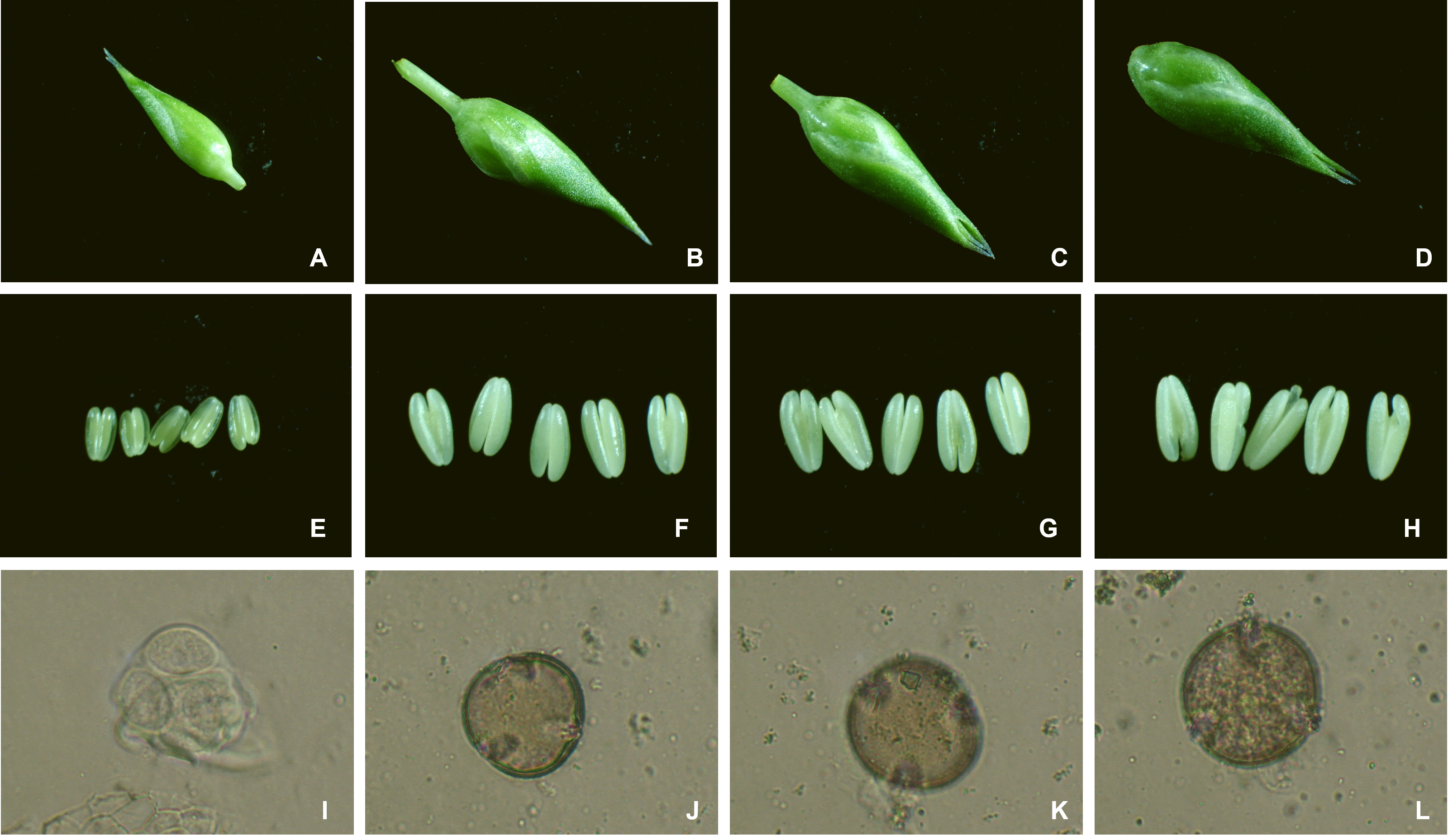
The induction of microspore cell division is an important step in androgenesis to produce doubled haploid plants. In Fytagoras’ research projects this process is followed closely.
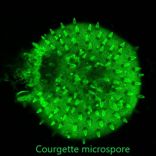
The movies shows a phase contrast time-lapse recording of approximately 48 hours, showing an embryogenically induced star-like microspore with a central nucleus and cytoplasmic strands radiating toward the exine wall like the spokes of a wheel. In the centrally located nucleus, two nucleoli are visible.
The nucleus moves toward the wall, transitioning from the star-like morphology to a cytoplasm-filled cell morphology. During this transition, the nucleus divides, and multiple nucleoli can be seen in each nucleus—three visible in the upper nucleus and two in the lower nucleus. It appears that two of the upper nucleoli are fusing, but later three are visible again, suggesting they may be moving around each other (the sudden jolt in the image is due to refocusing on the nucleus). At the end of the video, the two nucleoli in the lower right nucleus fuse into a larger single nucleolus.
The research strongly supports Fytagoras’ extensive experience in DH technology and successes in several recalcitrant crops like e.g. tomato, pepper, potato and many ornamental crops.